Cardiovascular System
In all multicellular organisms, living cells require nourishment, oxygen, and other important chemicals. Furthermore, waste or dangerous chemicals must be eliminated on a constant basis to ensure that tissues work properly. It is consequently necessary to have efficient pathways for transporting these chemicals to and from the cells. The cardiovascular system transports gasses, nutrition, hormones, and cellular waste throughout the body. It comprises mostly of three interconnected components: blood, the heart, and blood vessels.
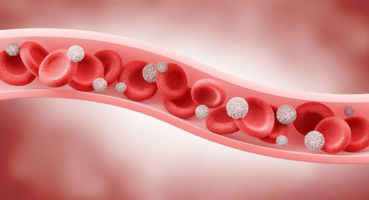
Blood
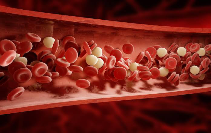
Blood is a connective tissue made up of blood plasma and other components. Blood is somewhat alkaline, with pH ranging from 7.3 to 7.4. It makes up 20-30% of extracellular fluid, accounting for 8% of total body mass. Blood serves the following vital functions:
- Transportation of gases like oxygen and carbon oxide, nutrients, hormones, heat and wastes.
- Regulation of pH, body temperature, and water content of cells
- Protection against blood loss through clotting and against disease
The Heart

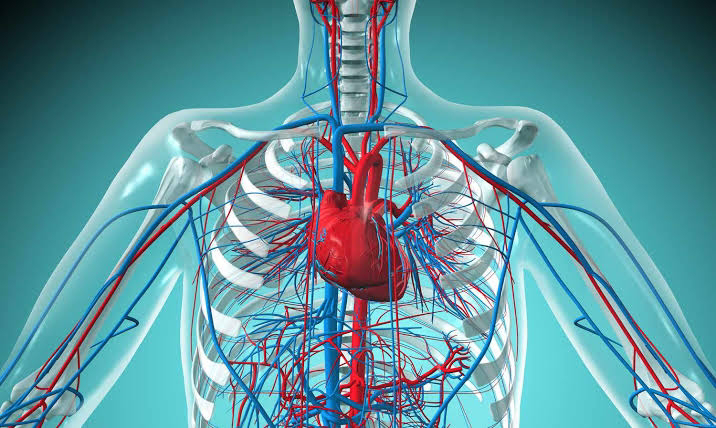
The heart is located in the thoracic cavity (chest), between the two lungs and slightly inclined toward the left lung. The pericardium is the membrane that surrounds and protects the heart; it is made up of an outer fibrous pericardium and an interior serous pericardium that form a double layer surrounding it. The pericardium cavity is a potential area filled with pericardial fluid that decreases friction between the two membranes. The wall of the heart is made up of three layers: epicardium, myocardium, and endocardium. The heart contains four chambers: two upper chambers, the right and left atria, and two inferior chambers, the right and left ventricles.
The right atrium receives blood from three veins: the superior vena cava, inferior vena cava, and the coronary sinus. It is divided from the left atrium by the interatrial septum. The Tricuspid valve directs blood flow from the right atrium to the right ventricle. It is also known as the right atrioventricular valve. The right ventricle receives blood from the right atrium. The interventricular septum separates it from the left ventricle, and it pumps blood through the pulmonary valve into the pulmonary trunk, a major artery that divides into the right and left pulmonary arteries. The left atrium receives oxygenated blood from the lungs via four pulmonary veins, two for each lung. The bicuspid valve allows blood to travel from the left atrium to the left ventricle. It is also known as the left atrioventricular valve. The left ventricle is the heart's thickest chamber. The aortic valve directs blood flow from the left ventricle to the ascending aorta. The left ventricle, which receives the most strain, has the thickest wall. It transports blood over long distances to all other regions of the body under high pressure.
Cord-like tendons known as chordae tendineae connect each atrioventricular valve flap. It connects the papillary muscles with the tricuspid and bicuspid valves. Chordae tendineae and papillary muscles keep the valve flaps closed.
Heart valves

The heart contains two pairs of valves. One set, the atrioventricular (AV) valves, keeps blood flowing in one direction between the atria and ventricles. The tricuspid valve is on the right side, whereas the bicuspid (or mitral valve) is on the left. Another pair of valves, known as the semilunar valves, ensures one-way passage from the ventricles to the arterial networks. The pulmonary valve is placed at the outlet of the right ventricle, while the aortic valve is located at the exit of the left ventricle. ejection of blood from the heart into the arteries while preventing backflow into the ventricles.
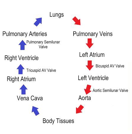
When the ventricles contract, pressure increases within the chambers. The semilunar valves open when the pressure in the ventricles surpasses the pressure in the arteries, allowing blood to flow from the ventricles into the pulmonary artery and aorta. As the ventricles relax, blood begins to flow back to the heart. The backflowing blood forces the semilunar valves to close tightly. There are no valves at the intersections of the venae cavae and the right atrium, or the pulmonary veins and the left atrium. Thus, as the atria contract, a little amount of blood flows backward from the atria into these veins. The left atrium receives oxygenated blood from the lungs. The right atrium recedes pulmonary trunk, which returns aorta to all organs throughout the body except right ventricle flowing into the pulmonary trunk, that carry blood To the left and right lungs. The left atrium receives oxygenated blood from the lungs. The left ventricles pump blood into the aorta. From the aorta to all organs in the body save the lungs. The right atrium receives deoxygenated blood returning from the systemic circulation. Blood pumped from the right ventricle enters the pulmonary trunk, which transports blood to the right and left lungs.
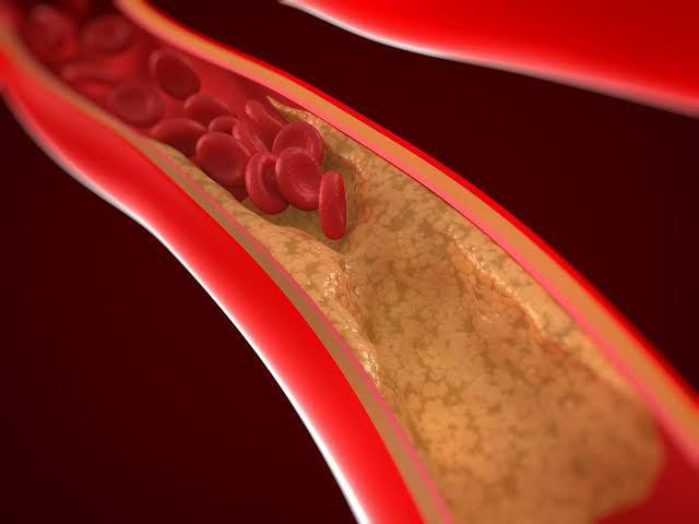
Coronary or cardiac circulation refers to the flow of blood via the blood arteries of the heart muscle (myocardium). Coronary arteries are the vessels that give oxygenated blood to the myocardium. Coronary veins are the vessels responsible for removing deoxygenated blood from the heart muscle. Coronary arteries deliver blood to the heart muscles (about 200 ml/min). These arteries have an extremely small diameter and can become clogged, resulting in a heart attack. A myocardial infarction, often known as a heart attack, happens when a part of the heart muscle dies from lack of oxygen. If a coronary artery becomes partially blocked, the individual may develop angina pectoris, which is characterized by radiating discomfort in the left arm. When a coronary artery becomes fully clogged, a heart attack occurs.
Even if the SA node fails to function effectively, the heart may continue to beat due to impulses generated by the AV node. However, the beat is slower, ranging between 40 and 60 minutes per minute. This problem can be treated by implanting an artificial pacemaker, which sends an electrical impulse to the heart every 0.8 seconds.
The heartbeat is the spontaneous regular contraction and relaxation of the heart to pump and receive blood to and from the body. When the heart beats, the atria contract first, followed by the ventricles. Then each chamber relaxes. The heart contracts, or beats, around 72 times each minute, with each beat lasting about 0.8 seconds.
The heart's intrinsic conduction system causes the atria and ventricles to contract rhythmically. The SA nodes commence the heartbeat and automatically send cardiac action potentials every 0.8 seconds, causing the atria to contract. When impulses reach the AV node, there is a tiny delay that permits the atria to finish contracting before the ventricles begin to contract.
Cardiac cycle
The cardiac cycle refers to the sequence of events that occur within a single heartbeat. It refers to the repeated contraction and relaxation of the heart. Systole refers to the contraction of the heart, whereas diastole refers to its relaxation. When the heart rate is 75 beats per minute, a cardiac cycle takes around 0.8 seconds. During atrial systole, the atria contract while the ventricles rest; the AV valves open. During ventricular systole, the atria relaxes and the ventricles contract; the AV valves are closed. During the relaxation period, which lasts about 0.4 second, the atria and the ventricles are both relaxed.
Heart sounds

During each cardiac cycle, four heart sounds are produced, but in a normal heart, only the first and second heart sounds are audible using a stethoscope. The first heart sound is low-pitched, soft, and rather protracted, similar to lub. The second heart sound has a higher pitch and is shorter and sharper, similar to dup. As a result, one typically hears "lub-dup-lub-dup-lub-dup". The first cardiac sound is connected with the closure of the AV valves (systole), while the second is associated with the closing of the semilunar valves (diastole).
Blood Vessels
The cardiovascular system contains three types of blood arteries. The arteries transport blood from the heart to various organs; the capillaries allow substances to exchange between the blood and bodily tissues; and the veins return blood from the tissues to the heart. The wall of the blood vessels consists of three layers: tunica interna (inner lining), tunica media (middle layer) and tunica externa (outer covering).
Arteries:
Arteries contain a thick, elastic coating that allows them to stretch while also absorbing strain. The arteries maintain pressure in the circulatory system. During ventricular systole, the heart pumps more blood into the arteries than flows out to the arterioles. The highly elastic arteries stretch temporarily to accommodate the excess amount of expelled blood. When the heart relaxes and temporarily ceases pumping blood into the arteries, the stretched artery walls passively recoil. An artery's wall has the same three layers as a conventional blood vessel, but it also has a thick muscular and elastic tunica media. Large arteries are called elastic (conducting) arteries, while medium-sized arteries are considered muscular (distributing).
As they reach their goal, arteries split into finer branches called arterioles. Unlike arteries, arteriolar walls feature a thick layer of smooth muscle that is well innervated by the sympathetic nerve. The smooth muscle is also susceptible to several local chemical variations. When the smooth muscle layer contracts, the vessel's circumference (and radius) shrinks, raising resistance and reducing blood flow through it. The narrowing of a vessel is known as vasoconstriction. In contrast, vasodilation refers to an increase in the radius of a vessel. Vasodilation causes less resistance and higher flow across the vessel. Arteries and arterioles transport blood away from the heart to the capillaries; capillaries connect arterioles to venules; and veins and venules return blood from the capillaries to the heart.
Veins:
A vein begins with the union of venous capillaries, which leads to the creation of venules, which eventually combine to create a vein. Venules move blood from capillaries to veins. Veins have thinner walls and a larger lumen, resulting in less barrier to blood flow. Veins have the same three layers as arteries, but their relative thicknesses differ. The tunica interna of veins is thinner than that of arteries, whereas the tunica medium of veins is significantly thinner, with less smooth muscle and elastic fibers. The tunica externa of veins is the thickest layer, made up of collagen and elastic fibers.
Many veins include valves, which are tiny folds of tunica interna that produce flaplike cusps. The valves point toward the heart. It lets blood to flow solely toward the heart while open and prevents blood from flowing backward when closed. Vein walls are thinner, so they can grow more easily.
Capillaries:

Each capillary is a tiny tube that is exceedingly narrow (usually less than 1 mm long) and has one-cell-thick walls made entirely of endothelium with a basement membrane. Capillary beds (networks of numerous capillaries) are found throughout the body, therefore a cut to any body tissue draws blood. The capillaries have a larger total cross-sectional area than the arteries or veins, hence the rate of blood flow (velocity) is lowest in the capillaries. Blood pressure is highest in the arteries but drops significantly as it passes through the capillaries. It's lowest in the veins. Capillaries are an essential component of the human circulatory system because they facilitate the passage of chemicals across their thin walls. Oxygen and nutrients, including glucose, flow from a capillary into the interstitial fluid that surrounds cells. Waste, like as carbon dioxide, diffuses into the capillaries. The relative consistency of interstitial fluid is completely dependent on capillary exchange.
Blood Pressure:

Blood pressure is defined as the arterial pressure of blood exerted on the artery wall with each heartbeat. Actually, blood pressure refers to the force exerted by blood on any unit area of the arterial wall. It is measured in millimeters of mercury (mmHg). The highest pressure exerted in the arteries as blood is discharged into them during systole. This is known as systolic pressure (an average of 120 mm Hg). The lowest artery pressure is known as diastolic pressure (average 80 mm Hg). Diastolic pressure occurs as the cardiac ventricles relax. Blood pressure is calculated as the ratio of systolic and diastolic pressures. A young adult's normal resting blood pressure is defined as 120 mm Hg over 80 mm Hg, or simply 120/80. The systolic pressure is higher, but the diastolic pressure is lower. Actually, blood pressure varies throughout the body. Blood pressure is highest in the aorta and lowest in the venae cavae. It is normal, however, to take the blood pressure in the brachial artery of the arm, which is usually 120/80. The difference between systolic and diastolic blood pressure is known as pulse pressure. When blood pressure is 120/80, the pulse pressure is 40 mm Hg (120 minus 80).
Circulatory routes
Blood vessels form circulatory channels that transport blood to certain organs in the body. The two primary circulatory channels are the systemic and pulmonary circulations.

The systemic circulation consists of the arteries and arterioles that transport oxygenated blood from the left ventricle to the systemic capillaries, as well as the veins and venules that return deoxygenated blood to the right atrium. The systemic circulation, which provides blood flow to all tissues of the body except the lungs, is also known as the larger circulation or peripheral circulation. The aorta is the biggest artery in the systemic circuit. The aorta is the body's biggest artery, with a diameter of 2-3 cm. All systemic arteries branch from the aorta. The largest veins are the superior and inferior venae cavae. The coronary (cardiac) circulation and hepatic portal circulation are two of the systemic circulation's subdivisions.


Comments
Post a Comment