During aerobic respiration, organisms use oxygen to oxidize respiratory substrates such as glucose, releasing energy for various activities. Catabolic processes also result in the release of carbon dioxide. As a result, oxygen must be continuously delivered to the cells, and the carbon dioxide created by the cells must be expelled. Respiration is the process of gaseous exchange in which cells absorb oxygen from the atmosphere and release carbon dioxide.
The process of respiration involves the following processes:
- Pulmonary ventilation or breathing - Exchange of air between the atmosphere and the alveoli of the lungs.
- Pulmonary (External) respiration - Exchange of oxygen & carbon dioxide between air in the alveoli and the blood within the capillaries.
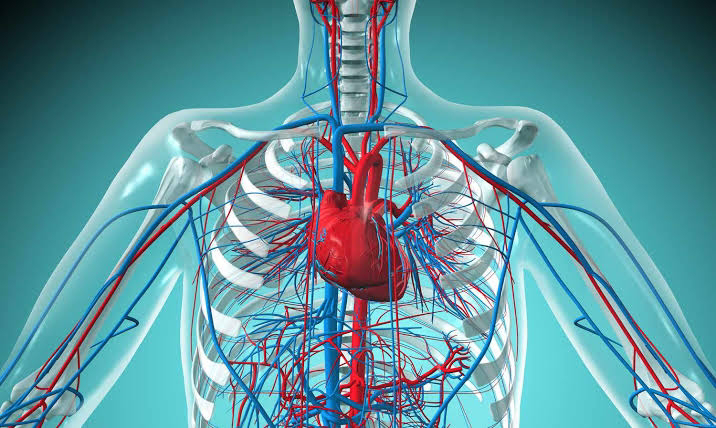
- Thee blood transports oxygen and carbon dioxide between the lungs and tissues.
- Internal respiration - Exchange of oxygen and carbon dioxide between the tissue cells and the blood.
Respiratory organs
The respiratory system exchanges gases (oxygen and carbon dioxide) between the atmosphere, blood, and tissue cells. In addition to gas exchange, the respiratory system regulates blood pH and contains receptors for the sense of smell. It also filters inspired air, generates vocal sounds, and expels small amounts of water and heat.
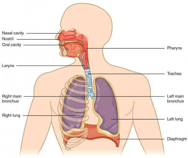
The respiratory system of human consists of-
- Nose
- Pharynx (throat)
- Larynx (voice box)
- Trachea (windpipe)
- Bronchi
- Lungs
The nose can be separated into two parts: exterior and interior. The external nose refers to the section of the nose that is visible on the face. The external nares, often known as nostrils, are two apertures on the undersurface of the external nose. The internal nose is a huge cavity known as the nasal cavity. The nose heats, moistens, and filters/cleans the air. The nasal cavity opens into the pharynx, which is the common channel for food and air. The pharynx (throat) is a muscular tube lined by a mucous membrane. It is separated into three anatomical regions: nasopharynx, oropharynx, and laryngopharynx (or hypopharynx).
The nasopharynx is the top section of the throat that enters into the nasal cavity. The larynx (voice box or sound box) is an airway that links the pharynx with the trachea. It contains the vocal cords (or vocal folds), which produce sound. The vocal cords are located right below the point at which the pharyngeal tract separates into the trachea and esophagus. The esophagus is a tube through which food goes into the stomach. Air enters the larynx through the gap between the vocal folds. The laryngeal orifice is referred to as the glottis. During swallowing, the glottis can be covered by a thin elastic cartilaginous flap known as the epiglottis to prevent food from entering the larynx. The laryngeal wall is made up of nine segments of cartilage.
Air generally enters the pharynx by the nose, although it can also enter via the mouth. Because the pharynx serves as a common conduit for food and air, reflex mechanisms seal off the trachea during swallowing, allowing food to enter the esophagus rather than the trachea. The esophagus remains closed unless when swallowing to prevent air from entering the stomach during breathing.
Beyond the larynx, the trachea separates (around the level of the fifth thoracic vertebra) into two main branches, the right and left primary bronchi, which enter the left and right lungs, respectively. When the principal bronchi enter the lungs, they divide into smaller bronchi known as secondary (lobar) bronchi, one for each lobe. The right lung has three lobes, while the left has two. The secondary bronchi continue to branch, generating smaller bronchi known as tertiary (segmental) bronchi, which further divide into bronchioles. Bronchioles branch frequently, eventually leading to smaller tubes known as terminal bronchioles. The vast branching from the trachea resembles an inverted tree and is known as the bronchial tree. Terminal bronchioles split into smaller branches known as respiratory bronchioles. The respiratory bronchioles penetrate deeper into the lungs. Respiratory bronchioles are further subdivided into two to eleven alveolar ducts.
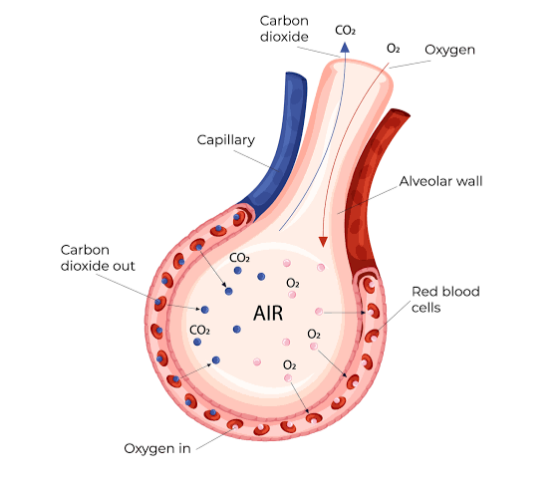
The incomplete cartilaginous rings support the trachea, primary, secondary, and tertiary bronchi, as well as the early bronchioles. Each terminal bronchiole produces a number of very thin, irregularly walled, and vascularized bag-like structures known as alveoli. An alveolus is a bag-like structure coated with simple squamous epithelium and supported by a thin elastic basement membrane; an alveolar sac is made up of two or more alveoli that have a common opening. The exchange of oxygen and carbon dioxide between the lungs' air spaces and the blood occurs via diffusion between the alveolar and capillary walls, which together form the respiratory membrane.
Humans have two lungs, which are placed in the thoracic or chest cavity. The outer chest wall (thorax) is made up of 12 pairs of curving ribs that are linked anteriorly by the sternum or breastbone and posteriorly by the thoracic vertebrae (backbone). The diaphragm is the floor of the thoracic cavity. It is a huge, dome-shaped membrane of skeletal muscle that divides the thoracic and abdominal cavities. The two lungs dangle freely within the cavity, connecting to the rest of the body only at one point where the lung's blood arteries and bronchus meet.
A pleural sac separates each lung from the thoracic wall. It is the thin fluid-filled gap located between each lung's two pulmonary pleurae. The pleura is a serous membrane. The pleural cavity refers to the interior of the pleural sac. The pleural surface secretes a serous fluid called intrapleural fluid, which lubricates the pleural surfaces as they slide past one other during respiration.
Mechanism and Breathing
Breathing or pulmonary ventilation consists of two phases: inspiration during which atmospheric air is drawn in and expiration by which the alveolar air is released out. The movement of air into and out of the lungs is carried out by creating a pressure gradient between the lungs and the atmosphere. Inspiration can occur if the pressure within the lungs (intrapulmonary pressure) is less than the atmospheric pressure, le., there is a negative pressure in the lungs with respect to atmospheric pressure. Similarly, expiration takes place when the intrapulmonary pressure is higher than the atmospheric pressure. The diaphragm and a specialised set of muscles (external and internal intercostals between the ribs) help in generation of such gradients.
Inspiration:
Inspiration (or inhalation) begins with the diaphragm contracting, increasing the volume of the thoracic chamber in the antero-posterior axis. During silent breathing, the inspiratory muscles (diaphragm and external intercostal muscles) contract to achieve inspiration. Before the start of inspiration, all respiratory muscles are relaxed. At the start of inspiration, the inspiratory muscles contract, expanding the thoracic cavity. The diaphragm is the principal inspiratory muscle. The relaxed diaphragm has a dome shape. When the diaphragm contracts, it lowers, expanding the capacity of the thoracic cavity by increasing its vertical dimension. The diaphragm contracts to account for 75% of the thoracic cavity's expansion during silent inspiration. When the external intercostal muscles contract, they raise the ribs and then the sternum upward and outward. It expands the thoracic chamber in the dorsoventral axis. The overall increase in thoracic volume induces a corresponding rise in pulmonary volume.
Before inspiration, at the conclusion of the previous expiration, intra-pulmonary (or intra-alveolar) pressure is equal to atmospheric pressure, indicating that no air is going into or out of the lungs. When the diaphragm and external intercostals compress during inspiration, the thoracic cavity expands, forcing the lungs to expand as well. As the lungs expand, the intrapulmonary pressure decreases. Because the intrapulmonary pressure is now lower than the atmospheric pressure, air enters the lungs via the pressure gradient from higher to lower pressure.
Expiration:
Breathing out, also known as expiration (or exhalation), occurs as a result of a pressure gradient, but in the opposite direction. The pressure in the lungs is larger than that of the surrounding atmosphere. Expiration begins as the diaphragm and inspiratory muscles relax. Relaxation of the diaphragm and intercostal muscles restores the diaphragm and sternum to their usual positions, lowering the thoracic volume and thus the pulmonary volume. The diaphragm resumes its natural dome-shaped position. This causes an increase in intrapulmonary (or intra-alveolar) pressure to slightly higher than atmospheric pressure, resulting in the ejection of air from the lungs, also known as expiration. Normal expiration during silent breathing, unlike inspiration, is a passive process in which no muscle contractions occur. Expiration is only active during strong breathing.
Exchange of Oxygen and Carbon dioxide
The objective of breathing is to deliver a constant supply of oxygen and to continuously remove carbon dioxide. During pulmonary and systemic gas exchange, oxygen and carbon dioxide diffuse from locations of higher partial pressures to those of lower partial pressure. A gas's partial pressure is the pressure it exerts in a mixture of gases. In pulmonary gas exchange, gases are exchanged between alveolar air and pulmonary blood capillaries through simple diffusion. Systemic gas exchange involves the exchange of gases between systemic blood capillaries and tissue cells.
Disorders of Respiratory system
Chronic obstructive pulmonary disease (COPD):
COPD is defined as any disorder that produces airflow obstruction, primarily due to constriction of airways. The primary symptoms are shortness of breath and a cough with sputum production. The most common COPDs include asthma, chronic bronchitis, and emphysema.
Asthma
Asthma is a chronic lung condition characterized by inflammation in the airway. Inflammation causes the airways to swell and become particularly sensitive to a number of stimuli. Airway obstruction can be caused by smooth muscle spasms in the walls of smaller bronchi and bronchioles, edema of the airway mucosa, increased mucus secretion, and/or airway epithelial damage. Symptoms may include difficulty breathing, coughing, wheezing, chest tightness, tachycardia, weariness, damp skin, and anxiety.
Emphysema
Emphysema is a condition characterized by abnormal air space growth caused by alveolar wall breakdown. The lungs also develop fibrosis and lose elasticity. With less surface area for gas exchange, oxygen diffusion across the injured respiratory membrane is diminished. It also increases the amount of air trapped in the lungs at the end of exhalation. Emphysema is typically induced by a prolonged irritation; the most common irritants are cigarette smoke, air pollution, and occupational contact to industrial dust.
Bronchitis
Branchitis is an inflammation of the bronchi in the lungs. There are two types of bronchitis: acute and chronic. Acute bronchitis (also known as a chest cold) typically causes a cough that lasts three weeks. It is a short-term inflammation of the bronchi in the lungs. More than 90% of the cases are caused by a viral infection. Chronic bronchitis is characterized as a productive cough that lasts three months or more per year for at least two years. Cigarette smoking is the primary cause of chronic bronchitis.
Pneumonia
Pneumonia is an inflammation of the lung's alveolar lining. Typical signs and symptoms include coughing, chest discomfort, fever, and dyspnea. The symptoms might range from mild to severe. Many factors influence how deadly pneumonia is, including the type of bacteria causing the infection, your age, and overall health. Pneumonia is typically caused by an infection with viruses or bacteria, while other microbes are less commonly involved. Bacteria are the leading cause of pneumonia. Streptococcus pneumoniae is the leading cause of bacterial pneumonia.
Tuberculosis
Tuberculosis is an infectious disease caused primarily by the bacteria Mycobacterium tuberculosis. Although various Mycobacterium species can cause tuberculosis, M. tuberculosis is the primary causal agent. Tuberculosis mostly attacks the lungs. Once the bacteria enter the lungs, they multiply and produce inflammation, prompting neutrophils and macrophages to travel to the area and absorb the germs, preventing their spread. If the immune system is not compromised, the germs remain latent for life; however, reduced immunity allows the bacteria to escape into the blood and lymph and infect other organs. Many people do not experience symptoms like as exhaustion, weight loss, lethargy, anorexia, a low-grade fever, night sweats, cough, dyspnea, chest pain, or hemoptysis until the disease has progressed.


Comments
Post a Comment